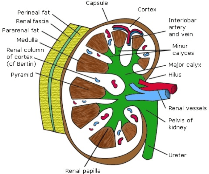
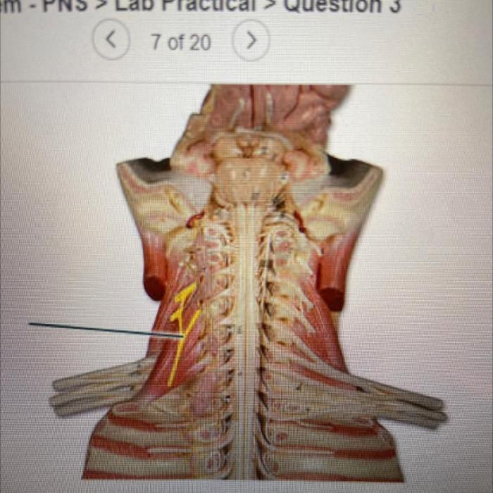
Identify the highlighted structure kidney – Embarking on a journey to identify the highlighted structure within the kidney, we unravel a fascinating tale of anatomy, function, and clinical significance. This structure, a cornerstone of the urinary system, plays a pivotal role in maintaining our body’s delicate balance.
Nestled within the renal parenchyma, this structure orchestrates a symphony of filtration, reabsorption, and secretion, ensuring the body’s optimal functioning. Its intricate histological features and cellular components form a harmonious ensemble that underpins its remarkable physiological capabilities.
Identify the Highlighted Structure Kidney

The kidney is a vital organ in the urinary system responsible for filtering waste products from the blood and producing urine. Identifying the highlighted structure within the kidney is crucial for understanding its function and potential clinical implications.
Anatomical Location
The highlighted structure is located within the renal medulla, the innermost region of the kidney. It is surrounded by the renal pelvis, which collects urine from the kidney, and the ureters, which transport urine to the bladder. The structure is also in close proximity to blood vessels that supply the kidney with oxygen and nutrients.

Structure and Function
The highlighted structure is a collecting duct, a network of tubules that collect urine from the nephrons, the functional units of the kidney. The collecting ducts merge to form larger ducts that ultimately empty into the renal pelvis. The collecting ducts play a crucial role in regulating the composition of urine by reabsorbing water and electrolytes from the filtrate.
Clinical Significance
Identifying the highlighted structure is clinically significant because abnormalities or diseases affecting this structure can impact kidney function. For example, damage to the collecting ducts can lead to impaired water reabsorption, resulting in conditions such as polyuria (excessive urine production) and dehydration.
Histological Analysis
Histologically, the collecting ducts are lined by a single layer of cuboidal or columnar epithelium. The cells are characterized by their pale cytoplasm and centrally located nuclei. The collecting ducts are surrounded by a thin layer of connective tissue.
Comparative Anatomy, Identify the highlighted structure kidney
The collecting ducts are found in the kidneys of all vertebrates. However, their structure and function may vary slightly among different species. For example, in mammals, the collecting ducts are longer and more complex than in non-mammalian vertebrates. This reflects the increased water conservation needs of mammals.
FAQ Resource
What is the primary function of the highlighted structure?
Filtration, reabsorption, and secretion, contributing to urine formation and maintaining electrolyte balance.
How does the structure’s anatomy influence its function?
Its intricate network of tubules and vessels facilitates efficient filtration and exchange of substances.
What clinical implications arise from abnormalities in this structure?
Dysfunction can lead to impaired kidney function, electrolyte imbalances, and various renal diseases.

